Bones In Leg Diagram : leg muscles diagram - Free Large Images - The foot bones shown in this diagram are the talus, navicular, cuneiform, cuboid, metatarsals and calcaneus.
byAdmin-
0
Bones In Leg Diagram : leg muscles diagram - Free Large Images - The foot bones shown in this diagram are the talus, navicular, cuneiform, cuboid, metatarsals and calcaneus.. The bones of the leg are the femur, tibia, fibula and patella. At the distal end of the femur, two rounded condyles meet the tibia and fibula bones of the lower leg to form the knee joint. The humerus and the femur are corresponding bones of the arms and legs, respectively. What does this suggest about mammals? The bones of your leg have roughened patches on their surfaces where muscles are attached.
The humerus and the femur are corresponding bones of the arms and legs, respectively. The majority of muscles in the leg are considered long muscles, in that they stretch great distances. Muscles and bones of the upper leg and pelvis; C) that they developed their bone structure independently of one another. He'll boost his body knowledge as he matches up the names of the bones with their proper places on the leg diagram.
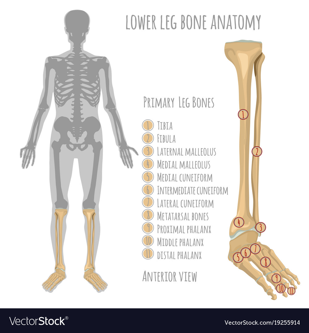
Lower leg bone anatomy Royalty Free Vector Image from cdn5.vectorstock.com The femur, or thigh bone, is the largest, heaviest, and strongest bone in the human body. Top suggestions for human leg bones diagram. The knee is a strong but flexible hinge joint. The majority of muscles in the leg are considered long muscles, in that they stretch great distances. Traditional methods to evaluate stress may be stressful to the bird. The basic bones of the human leg (image credit: The human leg, in the general word sense, is the entire lower limb of the human body, including the foot, thigh and even the hip or gluteal region. D) that the shape of the bones has less to do with the environment.
At the distal end of the femur, two rounded condyles meet the tibia and fibula bones of the lower leg to form the knee joint.
Want to learn more about bones? An electrical wiring diagram can be as simple as a diagram demonstrating how to set up a fresh swap with your hallway. The foot bones shown in this diagram are the talus, navicular, cuneiform, cuboid, metatarsals and calcaneus. Most of the animals have the same bones, although some are shaped differently and placed in different positions. , knee, and leg pain helps you understand how trigger points form and where to search for them. The foot bones shown in this diagram are the talus, navicular, cuneiform, cuboid, metatarsals and calcaneus. Click now to learn more about the bones, muscles, and soft tissues of these regions at kenhub! C) that they developed their bone structure independently of one another. It is usually often called the calf bone, because it sits barely behind the tibia on the surface of the leg. The human leg, in the general word sense, is the entire lower limb of the human body, including the foot, thigh and even the hip or gluteal region. Master leg and knee anatomy using our topic page. These leg muscle diagrams show you the major muscles of the human leg. The majority of muscles in the leg are considered long muscles, in that they stretch great distances.
What does this suggest about mammals? Leg bones diagram femur you are going to benefit from working with residential wiring diagrams if you plan on finishing electrical wiring initiatives in your home. These leg muscle diagrams show you the major muscles of the human leg. Color the leg on the left side. Top suggestions for human leg bones diagram.
4.1-2 shows the rabbit skeleton. The bones used in this ... from www.researchgate.net The foot bones shown in this diagram are the talus, navicular, cuneiform, cuboid, metatarsals and calcaneus. The foot bones shown in this diagram are the talus, navicular, cuneiform, cuboid, metatarsals and calcaneus. The foot bones shown in this diagram are the talus, navicular, cuneiform, cuboid, metatarsals and calcaneus. At the distal end of the femur, two rounded condyles meet the tibia and fibula bones of the lower leg to form the knee joint. Posted on january 20, 2015 by admin. Editor · aug 13, 2017 ·. The femur, or thigh bone, is the largest, heaviest, and strongest bone in the human body. The human leg, in the general word sense, is the entire lower limb of the human body, including the foot, thigh and even the hip or gluteal region.
The bones of your leg have roughened patches on their surfaces where muscles are attached.
Traditional methods to evaluate stress may be stressful to the bird. Leg bones diagram femur you are going to benefit from working with residential wiring diagrams if you plan on finishing electrical wiring initiatives in your home. When you stand or walk, all the weight of your upper body rests on them. What does this suggest about mammals? Related posts of leg bones anatomy diagram structure of anatomy leg and foot. Editor · aug 13, 2017 ·. The majority of muscles in the leg are considered long muscles, in that they stretch great distances. The humerus and the femur are corresponding bones of the arms and legs, respectively. Learn vocabulary, terms and more with flashcards, games and other study tools. The foot bones shown in this diagram are the talus, navicular, cuneiform, cuboid, metatarsals and calcaneus. When your muscles contract, they pull the bone they're. At the distal end of the femur, two rounded condyles meet the tibia and fibula bones of the lower leg to form the knee joint. Mcqs on leg bones for neet.
Growth of the long bones in a juvenile knee joint (the femur is located proximally the anterior muscular pouch on the knee joint, anchored by the quadriceps tendon and patellar tendon on the distal anterior femoral surface (see diagram below). Bones pain hand and arm bones diagram. He'll boost his body knowledge as he matches up the names of the bones with their proper places on the leg diagram. When you stand or walk, all the weight of your upper body rests on them. While their parts are similar in general, their structure has been adapted to differing functions.
Dem Bones, Dem Bones: The Skeleton from www.susaningraham.net He'll boost his body knowledge as he matches up the names of the bones with their proper places on the leg diagram. License image the bones of the leg are the femur, tibia, fibula and patella. At the distal end of the femur, two rounded condyles meet the tibia and fibula bones of the lower leg to form the knee joint. Human anatomy diagrams show internal organs, cells, systems, conditions, symptoms and sickness information and/or tips for healthy living. The bones of the leg are the femur, tibia, fibula and patella. Top suggestions for human leg bones diagram. He leg's main function in the human is for locomotion and support of the rest of the body. While their parts are similar in general, their structure has been adapted to differing functions.
Bones pain hand and arm bones diagram.
Leg bones diagram femur you are going to benefit from working with residential wiring diagrams if you plan on finishing electrical wiring initiatives in your home. The foot bones shown in this diagram are the talus, navicular, cuneiform, cuboid, metatarsals and calcaneus. The femur, or thigh bone, is the longest, heaviest, and strongest bone in the body. While their parts are similar in general, their structure has been adapted to differing functions. The knee is a strong but flexible hinge joint. Posted on january 20, 2015 by admin. The foot bones shown in this diagram are the talus, navicular, cuneiform, cuboid, metatarsals and calcaneus. Drawing of the muscles in legs and feet wellcome v0008871.jpg 2,469 × 2,970; The human leg consists of 8 bones, 4 per leg. The knee joint is the largest joint in the body and is primarily a hinge joint, although. Related posts of leg bones anatomy diagram structure of anatomy leg and foot. The femur, or thigh bone, is the largest, heaviest, and strongest bone in the human body. The bones of your leg have roughened patches on their surfaces where muscles are attached.